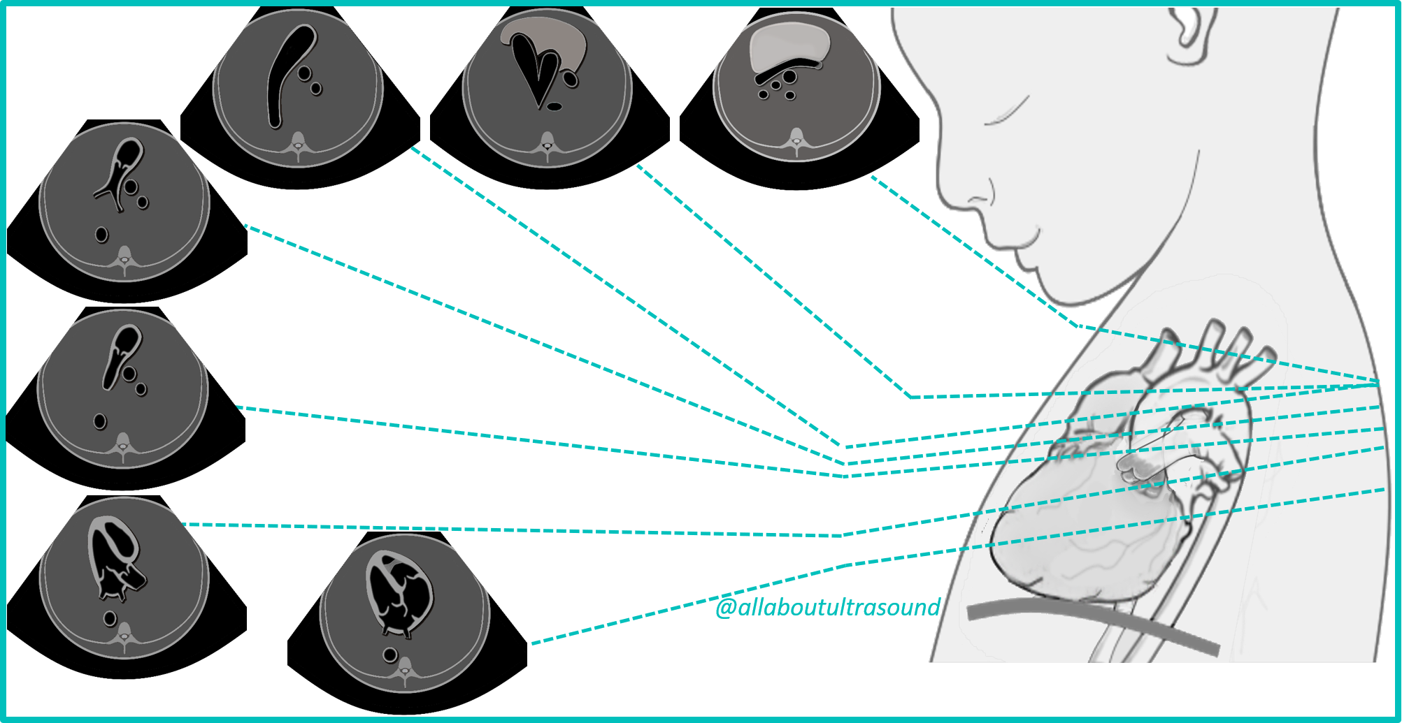
Fetal Echo Views
13-view sweep sequence
1
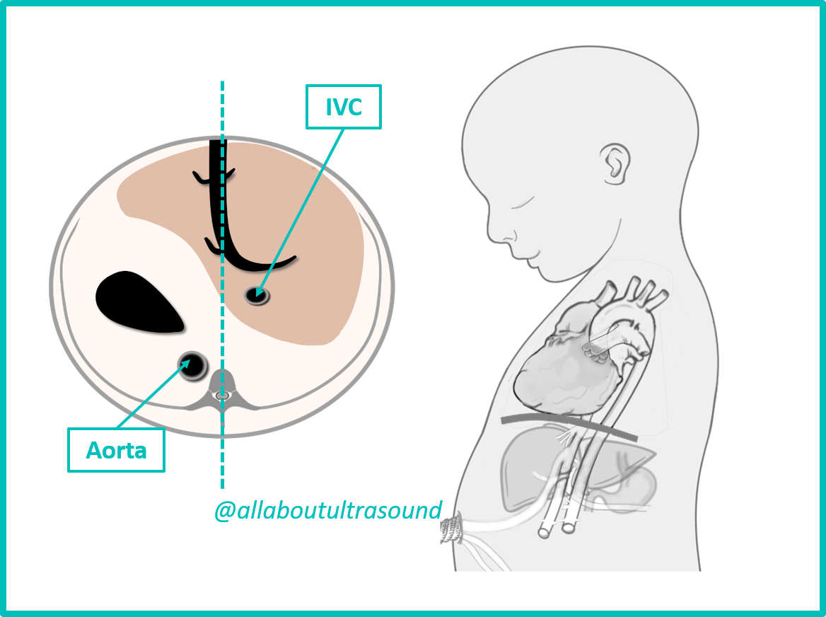
Abdominal Situs View
SitusThe first step in fetal cardiac evaluation. Confirms normal situs solitus — stomach on left, liver on right, aorta left of spine, IVC right of spine. Situs abnormalities are strongly associated with complex congenital heart disease.
View Reference Images
··
Anatomy Diagram
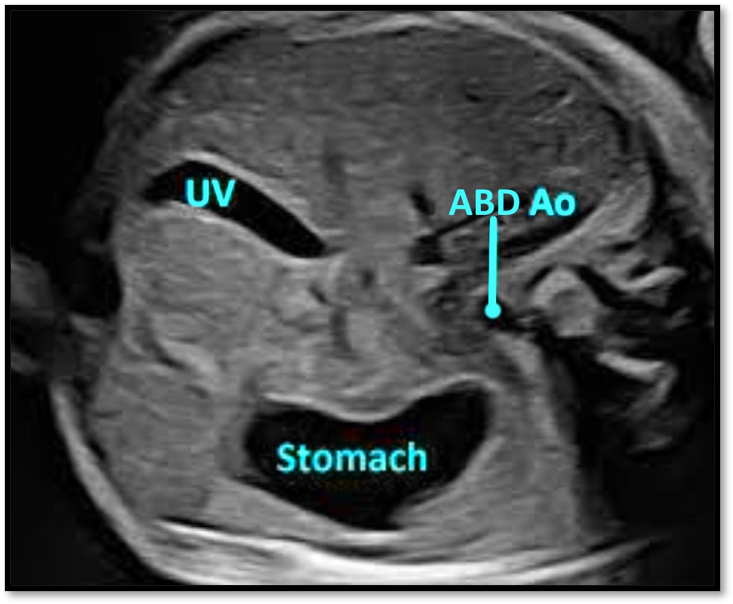
Clinical Echo Image
Structures to Identify
- Stomach (left)
- Liver (right)
- Descending aorta (left of spine)
- IVC (right of spine)
- Umbilical vein
- Spine (posterior)
Normal Findings
- Stomach bubble on LEFT side of fetus
- Aorta to LEFT of spine, IVC to RIGHT
- Liver on right, stomach on left
- Umbilical vein entering liver anteriorly
Scanning Technique
Transverse view of fetal abdomen at level of stomach. Identify spine posteriorly. Confirm stomach on left and aorta/IVC positions relative to spine.
Patient Positioning
Mother supine or left lateral tilt; obtain true transverse cardiac cut perpendicular to fetal spine
Doppler
Color Doppler to confirm aorta (pulsatile) vs. IVC (venous) positions relative to spine.
Common Pitfalls
- Fetal position may make left/right orientation confusing — always reference spine first
- Absent stomach may indicate esophageal atresia or diaphragmatic hernia
Red Flags / Abnormal Findings
- !Stomach on RIGHT (situs inversus or heterotaxy)
- !Stomach absent (esophageal atresia, CDH)
- !Aorta and IVC on same side (asplenia/polysplenia)
- !Midline stomach (heterotaxy)
Clinical images © All About Ultrasound, Inc. / iHeartEcho™. Educational use only.